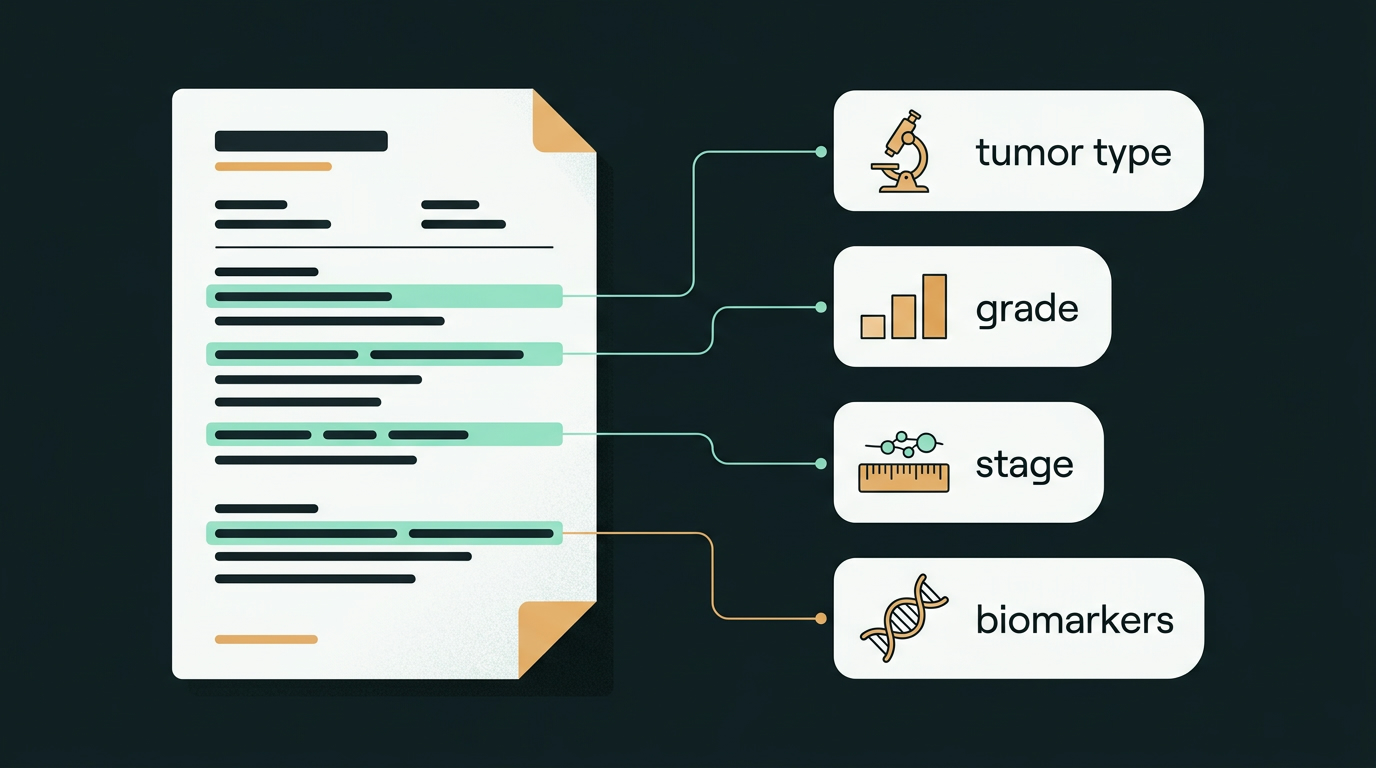
A pathology report is often the most important document in a cancer diagnosis — and one of the hardest to read. It is written by doctors, for doctors, in a compressed language of abbreviations, percentages, and codes. Many people now see it in a hospital app or patient portal before anyone has had a chance to explain it, and a page full of unfamiliar words can be frightening.
This guide walks through the parts of a typical report and what the most common terms mean. It will not tell you what your report means for you — that depends on many things only your treating team knows. But understanding the vocabulary can turn a confusing document into a list of good questions, and make the conversation with your doctor far more useful.
What is a pathology report?
When a biopsy or surgery removes tissue, it is sent to a pathologist — a doctor who specialises in diagnosing disease by examining cells and tissue under a microscope. The pathologist's report gives the definitive diagnosis of cancer and provides much of the information used to stage it and plan treatment.
A standard report is usually sent to your doctor within about 10 days of the biopsy or surgery, because the tissue needs several days of processing. Additional tests, such as some biomarker or genetic tests, may be reported separately and take longer.
The main sections
- Patient and specimen information — your details, the date, which part of the body the tissue came from, and how it was obtained (for example, a needle biopsy or surgical removal).
- Gross (macroscopic) description — what the pathologist sees with the naked eye: the size, weight, and appearance of the tissue, and how many lymph nodes were received.
- Microscopic description — what the cells look like under the microscope, including the type of cancer, its grade, the margins, and the lymph nodes.
- Diagnosis — the pathologist's summary. If you read only one section, this is usually the most important one.
- Comments — optional notes, such as unusual findings, tests still pending, or extra tests the pathologist recommends.
Key terms, decoded
Cancer type
The name tells you which kind of cell the cancer started in. "Carcinoma" means a cancer that began in the cells lining organs and glands; "adenocarcinoma" is a carcinoma that began in gland-forming cells. "Invasive" means the cancer has grown beyond the layer where it started. "In situ" means the abnormal cells have not grown into nearby tissue.
Grade (G1, G2, G3)
Grade describes how abnormal the cancer cells look compared with normal cells. Grade 1 (well differentiated, low grade) cells look closest to normal and tend to grow more slowly. Grade 2 is intermediate. Grade 3 (poorly differentiated, high grade) cells look very abnormal and tend to grow faster. "GX" means the grade could not be assessed. Grade is not the same as stage.
Margins
After surgery, the pathologist checks the edges of the removed tissue. A negative (clear) margin means no cancer cells were seen at the edge, suggesting the tumour was fully removed. A positive (involved) margin means cancer cells reached the edge, suggesting some may remain. Your surgeon and oncologist will explain whether anything further is needed.
Lymph nodes
Lymph nodes are small glands that can trap cancer cells spreading from a tumour. Reports often give a fraction such as "2/3 positive," meaning cancer was found in 2 of the 3 nodes examined. "Sentinel node" refers to the first node (or nodes) a tumour drains to, which surgeons often check first.
TNM stage
The TNM system is the most widely used way of describing how far a cancer has spread:
- T (tumour) — the size and extent of the main tumour, from T1 to T4. TX means it could not be measured.
- N (nodes) — whether nearby lymph nodes contain cancer. N0 means none; N1 to N3 mean increasing involvement.
- M (metastasis) — whether the cancer has spread to distant parts of the body. M0 means no distant spread was found; M1 means it has spread.
Small letters in front matter too. A "p" (as in pT2) means the stage was measured by the pathologist on tissue removed at surgery. A "c" means a clinical stage, estimated from examination, scans, and biopsies. A "y" (as in ypT2) means the stage was measured after treatment given before surgery. The exact meaning of each number differs between cancer types, so ask your team what your T and N numbers mean for your specific cancer.
Biomarkers
Biomarkers are genes, proteins, and other substances in the cancer that can help guide treatment. Which ones are tested depends on the type of cancer. Common examples include:
- ER and PR (oestrogen and progesterone receptors) in breast cancer — if present, hormones can drive the cancer's growth, and hormone therapy may be an option.
- HER2 in breast and some other cancers — scored 0, 1+, 2+, or 3+. See What Does HER2 Status Mean for Your Breast Cancer Treatment?
- Gene changes such as EGFR in some lung cancers, found by tumour (genomic) testing. Some changes match a targeted treatment; others are listed as "variants of unknown significance" and are generally not used for treatment decisions.
Tumour testing looks for changes in the cancer cells themselves, which are not passed on to family members. That is different from inherited (germline) testing, which looks for changes you were born with. If an inherited change is suspected, your team may suggest genetic counselling.
A worked example (fictional)
Here is how these pieces fit together on a short, made-up breast cancer report. It is not a real patient's report and is shown only to illustrate the terms above.
ER 90% PR 60% HER2 IHC 3+.
pT2 pN1.
Margins: negative.
Sentinel nodes: 2 / 3 positive.
The cancer cells look moderately abnormal — an intermediate grade.
The cells have hormone receptors, so hormone therapy is likely to be part of the treatment discussion.
Measured on surgical tissue. In breast cancer, T2 means a tumour more than 2 cm and up to 5 cm across; N1 means cancer in a small number of nearby lymph nodes.
Fictional example for learning only. Real reports vary in layout and detail.
What a pathology report cannot tell you on its own
It is natural to search each term and try to work out what lies ahead. Please be gentle with yourself here. A pathology report is one important piece of a larger picture that also includes scans, blood tests, your general health, and your own wishes. Two people with similar-looking reports may be advised quite differently, for good reasons.
Also note that a cancer keeps the stage it was given at diagnosis. If things change later, that new information is added, but the original stage stays the same.
Questions to ask your care team
- "What type of cancer is this, and what is its grade?"
- "What is the stage, and was it measured clinically or on surgical tissue?"
- "Were the margins clear? Were any lymph nodes involved?"
- "Which biomarker or genetic tests were done, and are any results still pending?"
- "How do these results affect my treatment options?"
- "Can I have a copy of my report?"
You can also ask whether the pathologist is available to talk through the findings. If you would like a second pathology opinion, discuss it with your doctor first: it usually needs the original slides or tissue block, which can be requested from the hospital or laboratory that made the diagnosis.
Frequently asked questions
What is the difference between tumour grade and stage?
Grade describes how abnormal the cancer cells look under the microscope. Stage describes how large the tumour is and how far the cancer has spread. They are separate measures, and doctors use both, along with other information, to plan treatment.
What does a positive margin mean?
A positive (involved) margin means cancer cells were seen at the edge of the tissue that was removed, which suggests some cancer may remain. A negative (clear) margin means no cancer cells were seen at the edge. Your surgeon and oncologist will explain whether any further treatment is needed.
What does the small p in pT2 or pN1 mean?
A small p means the stage was measured by the pathologist on tissue removed during surgery (pathological stage). A small c means a clinical stage, estimated from examination, scans, and biopsies before treatment. A small y means the stage was measured after treatment given before surgery.
Can I understand my prognosis from the pathology report alone?
No. The report is one important piece of information. Your treating team combines it with scans, blood tests, your general health, and other factors to plan treatment and discuss what to expect. Ask them to go through your report with you.
Can I get a second opinion on my pathology?
Yes. Talk to your doctor first. A second pathology opinion usually requires the original slides or tissue block, which can be requested from the hospital or laboratory that made the diagnosis. Some specialist centres offer this review.
Sources
- National Cancer Institute. Pathology Reports. https://www.cancer.gov/about-cancer/diagnosis-staging/diagnosis/pathology-reports-fact-sheet
- National Cancer Institute. Tumor Grade. https://www.cancer.gov/about-cancer/diagnosis-staging/diagnosis/tumor-grade
- National Cancer Institute. Cancer Staging. https://www.cancer.gov/about-cancer/diagnosis-staging/staging
- American Cancer Society. Cancer Staging. https://www.cancer.org/cancer/diagnosis-staging/staging.html
- National Cancer Institute. Biomarker Testing for Cancer Treatment. https://www.cancer.gov/about-cancer/treatment/types/biomarker-testing-cancer-treatment
- National Cancer Institute. Hormone Therapy for Breast Cancer. https://www.cancer.gov/types/breast/breast-hormone-therapy-fact-sheet
Last reviewed: November 1, 2026
Medical guidance changes; check the latest official recommendations.
This website provides general educational information only. It is not medical advice, diagnosis, or treatment, and it does not create a doctor-patient relationship. Consult a qualified healthcare professional about your own condition. In an emergency, call 997.